lambda monocut ladder (New England Biolabs)
93
Structured Review
New England Biolabs
lambda monocut ladder
Lambda Monocut Ladder, supplied by New England Biolabs, used in various techniques. Bioz Stars score: 93/100, based on 47 PubMed citations. ZERO BIAS - scores, article reviews, protocol conditions and more
https://www.bioz.com/result/lambda monocut ladder/product/New England Biolabs
Average 93 stars, based on 47 article reviews
lambda monocut ladder - by Bioz Stars,
2026-06
93/100 stars
Images
1) Product Images from "Synthetic genomic reconstitution reveals principles of mammalian Hox cluster regulation"
Article Title: Synthetic genomic reconstitution reveals principles of mammalian Hox cluster regulation
Journal: bioRxiv
doi: 10.1101/2021.07.07.451065
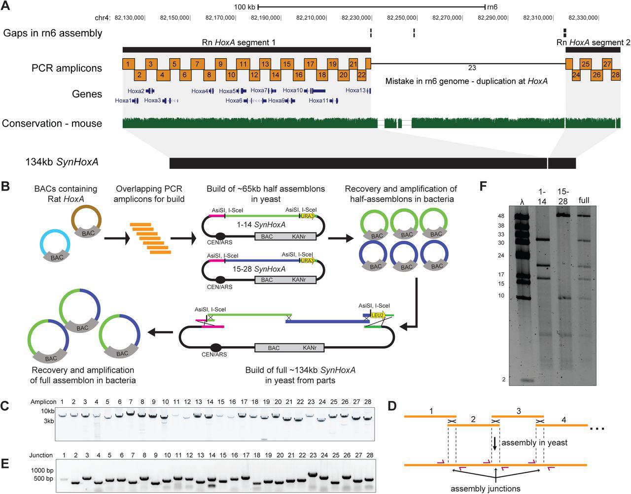
Figure Legend Snippet: (A) Layout of rat HoxA locus in the rn6 genome assembly. The rn6 genome includes an erroneous duplication at the HoxA locus between gaps in the assembly. The SynHoxA assemblon sequence is based on bringing together the two ‘separate’ RnHoxA segments. The sequence was segmented into 28 ∼5kb PCR amplicons with terminal homology of ∼200bp to adjacent amplicons. Conservation to the mouse genome is depicted using the multiz track from the UCSC genome browser. (B) Schematic depicting the assembly workflow for the 134kb SynHoxA assemblon. BACs containing Rat HoxA were used as PCR template to generate 28 segments tiling the entire HoxA locus. These segments were co-transformed into yeast with appropriate linkers and assembly vector to build two ∼65kb half assemblons into centromeric yeast-bacteria shuttle vectors. These half assemblons are recovered to bacteria and amplified. Full 134kb assemblon was built from half assemblons after releasing them from the vector using terminal restriction enzymes ( AsiSI ) and transforming into yeast. Full assemblon was then recovered from yeast into bacteria for amplification and verification. (C) Agarose gel of the 28 PCR amplicons that tile the 134kb SynHoxA assemblon. (D) Strategy to PCR-screen yeast colonies derived from assembly experiments. Primers (red arrows) span assembly junctions and test presence/absence of amplicons in many yeast colonies. Reproduced from ref with permission from authors. (E) Agarose gel showing one yeast colony carrying the full 134kb SynHoxA assemblon verified manually for the presence of all assembly junctions, using the strategy outlined in panel D. (F) Half and Full 134kb SynHoxA assemblon BACs purified from E.coli were digested with PvuI and separated using field inversion gel electrophoresis (FIGE). Lambda monocut ladder sizes are indicated in kb. Band sizes correspond to expected fragments.
Techniques Used: Sequencing, Transformation Assay, Plasmid Preparation, Amplification, Agarose Gel Electrophoresis, Derivative Assay, Purification, Nucleic Acid Electrophoresis

Figure Legend Snippet: (A) Layout of rat HoxA locus from the rn6 genome assembly depicting genes, Rn HoxA cluster segments in black and previously identified distal enhancers in purple. The Enhancers+SynHoxA assemblon sequence is made by stringing all the enhancers directly upstream of the SynHoxA assemblon sequence. Conservation to mouse genome is depicted using multiz track from the UCSC genome browser. (B) PCR amplicons tiling enhancer sequences were generated from Rat HoxA BACs and co-transformed into a yeast strain containing the 134kb SynHoxA assemblon with a gRNA vector targeting the left terminus of the 134kb assemblon. The enhancer PCR amplicons were used to repair this break, resulting in the construction of the 170kb Enhancers+SynHoxA assemblon. Assemblon was recovered into bacteria for amplification and verification. (C) Agarose gel of the 8 PCR amplicons containing enhancer sequences. (D) Agarose gel showing one yeast colony tested for the presence of novel enhancer assembly junctions and with primers spanning 134kb SynHoxA . (E) 134kb and 170kb assemblon BACs purified from E.coli were digested with PvuI and separated using FIGE. Lambda monocut ladder sizes are indicated in kb. Band sizes correspond to expected fragments.
Techniques Used: Sequencing, Generated, Transformation Assay, Plasmid Preparation, Amplification, Agarose Gel Electrophoresis, Purification

Figure Legend Snippet: (A) Schematic of assembly strategy for 130kb RAREΔ SynHoxA and 166kb Enhancers + RAREΔ SynHoxA . Nature of the RARE mutations is shown on the right. RAR binding data comes from previously published reports. (see Methods) (B) Sanger sequencing traces confirmed precise CRISPR editing of RAREs in yeast. (C) SynHoxA assemblon BACs purified from E.coli were digested with PvuI and separated using FIGE. Lambda monocut ladder sizes are indicated in kb. Bands correspond to expected fragment lengths. (D) Sequencing data of assemblon BACs purified from E. coli aligned to a custom mm10 reference genome. Positions of the enhancers and protein coding genes are shown in black.
Techniques Used: Binding Assay, Sequencing, CRISPR, Purification